“The Body in the Concrete”- Concrete Casket 101.

So last time, I expressed my concern on how the Hong Kong Police and Firemen had mistreated the crucial trace evidence, namely the concrete casket. And I also opposed their methodology. Some of the readers thought that police should be more experienced on handling these cases than me, and also thought that the law enforcement units had made this judgment after chains of thorough thinking.

I supposed I am not in a good position to comment further on how their approach was when they were at the scene. After all, I was not at the crime scene in person. Also, readers state that that was indeed the raw differences between theories, archaeology and the reality. I am only wishing to use the following space to replied to three of the main questions raised by the readers. I have also cited the Los Angeles Medical Examiners case report, in hope of the M.E. would be able to shed some lights on the questions the readers made from their study and research.

Question 1: The size of the concrete casket is too huge! May be they are not scanned because the law enforcement was not able to transfer them for scanning?

If you ever watched any crime shows on TV (of course, Bones is a good example. Everyone will yell “back to the lab!”), you will see they are always able to transfer whichever evidence they found back to the lab before further analyzing. Reality, not so much. This is how Ground Penetrating Radar comes into play. In archaeology, Ground Penetrating Radar (GPR) is used to detect and reflect any buried artifacts, monuments, archaeological sites. It is especially handy when archaeologists are about to look for hidden burial sites and buried remains. GPR allows noninvasive examination, and very helpful for experts and scientists to learn about the structure of the hidden architectures and bodies. Furthermore, size of a GPR is only about the size of a vacuum. Some companies even invented the GSSI Mini, which is about the size of a laptop for carrying scientists to have easy access in the field. All GPR and GSSI Mini come with a monitor, and very easy to connect to the laptop. That said, it is easy to document digitally the detected images. One may use slightly more time on using the GPR before stepping or unfold the crime scene, yet save the team and resources from doing extra and additional steps and procedures in the later investigation. In this case, that would be the suspicious broken palm, posture of the body, etc.

Question 2: It was mandatory to crack the concrete casket open, as the body was decomposing already.

News and police report claimed that the concrete casket was dried by the time of discovery. Yet, concrete would not dry but only cure and hardened. The hardening and curing of concrete, in other words, does not come from the evaporation of water from the chemical composition of the concrete. Rather the water molecules have transformed, merged and bonded together with the concrete particles as part of their chemical structure. An experiment pointed out that the mass of concrete before hardening/ curing is about the same with after [1]. The only slight difference between the mass was from the evaporation of the water on the concrete surface that with no cover. Last time, I have also mentioned that concrete is relatively porous. When cement hardens, it means that the water molecules and air molecules have filled in all those pores. This type filling makes concrete looks strong but indeed not. That said, it is relatively soft inside, while the outside of the concrete looks hard.

And for the body that was covered by the concrete, the decomposition of it liquefies from inside to outside, and all the decomposition was triggered by the enzymes in muscles. During the hardening process of the concrete, since it is a exothermic reaction (i.e. it releases heat in the whole process), the interior of the concrete casket would reach 175F in the first few days, which results an acceleration in the decomposition rate. After curing, the concrete becomes a good insulator that blocked the air and heat to reach the body, and thus successfully decrease the rate of decomposition again.

During the stage of decomposition, body liquids (any liquid in the body, you name it :)) would leak out of the body. Normally, as in general when a body is exposed to air, atmospheric air would help evaporate liquids and water. However, when a body like the one in this case is being buried in a concrete casket, all the fluid is trapped in the casket, and at the end turned the soft tissues into a mush. At the end, fluids would leak outside the casket, or concrete casket. This is not only something visual but also would give a strong odor. Evenly so, it does not mean an invasive act should be taken to the casket. Keep in mind that bodies starts breaking down the moment the heart stopped beating.

Question 3: Readers think that using merely textbook archaeology, i.e. using brush in the act, has not thoroughly considered the scenario at scene.

To be honest, this is some attitude that forensic scientists should have and maintain all along. In the forensic field, a lot of the tools we use are indeed very creative. For instance, you would find ladles on the autopsy table to scoop out fluid (for example inflammatory fluids in lungs) during autopsy; also would find those big stock pots in decomp bodies autopsy room for forensic anthropologists to do maceration. All these kitchenware is used with one and foremost premise: will not affect the quality of the collected evidences, or would not contemning evidences.

Los Angeles Medical Examiner Office claimed that there were only 5 cases of concrete casket located, till 2008, in the past 18 years in a report. They also stated in the report that, though cement and concrete affected the calculation or estimation of accurate postmortem interval, at the sam time they welly preserved all trace evidences [2]. In these 5 recorded cases, medical examiners were taken things slow, and excavate the bodies layer by layer in order to estimate the cause of death and time of death. Among all, LA medical examiners also indicated the frequent application of metal detectors and radiography in order to pinpoint the posture of bodes, and location. Sledgehammer and chisel are only implemented in a very much later stage, or only when they are sure it would not damage the body.

Back to the discussion, should we use heavy tools like sledgehammer and chisel, or only brush? Both. The foremost premise here is to not damaging the evidence. Only use heavy tools when the remains inside are well-documented. Also, the methodology with chisel should be go horizontally instead of vertically in order to reveal the context of the casket and the body.

Sad but true, concrete casket or related research is not commonly seen and discussed in the academia. These caskets can only open when all the conditions and situations are well-documented. In delicate crime scene like this one, officials should prioritize the preservation of crime scene in front of investigation just yet.


Remarks:

[1] Lesson 5: So, You Think Concrete Dries Out? (n.d.). Retrieved April 11, 2016, from

[2]
Toms, C., Rogers, C. B., & Sathyavagiswaran, L. (2008).
Investigation of Homicides Interred in Concrete—The Los Angeles
Experience. J Forensic Sci Journal of Forensic Sciences, 53(1), 203-207.
doi:10.1111/j.1556-4029.2007.00600.x

Forensics Daily #10: Estimating the Time of Death from Drowning and Remains Found in Water

Q: Is it possible to tell if victims died from drowning and the time of death of remains found in water?
A: yes, for sure there are several ways to tell if the victims were drowned. One of the ways, that is also help determine the time of death of remains found in water is to evaluate the diatom level.
Diatom refers to a diverse group of algae. It has no specific structure or characteristics as it is consider a general term of groups of algae that inhabit the remains. Yet, the migration of diatoms, meaning the sequences of diatoms in the body should tell the time of the body in the water by calculating the schedule.
The same applies to the drowning victim too! Yet, during the drowning process, victim should also have inhaled some of the fresh water (only in fresh water will find diatoms!). So in the lungs, ME should be able to find fresh water and diatoms for calculation, as well as backward calculating the appearance of diatoms in order to work out the time of death.
P.s. Diatoms belongs to the Forensic limnology department, which is a sub-discipline of forensic botany, fyi : ) !

Forensics Daily #9: Dead Wives Tales

Q: are the dead wives tales true at all? Meaning, our nails and hair do grow after we died ?

A: Yes and No. Yes, because it does look like it grows, like illusion. No, because it is NOT REALLY GROWING.. Both hair and nails look like they grow after the heart stopped working because of the skin surrounding them are dehydrated. They shrink, and thus make it appear to be longer. Funeral homes sometimes will moisturize the bodies when they are doing the prep to counteract this.

Other than the hair and nails, the chin also dries out, and pulling towards the back of a skull, thus looks like more prominent. So does the goosebump effect, because of the contraction around the hair muscle (remember we talked about in the last few q&a, after we died, our muscles are not able to relax, not unable to contract!).

These all biological responses give the sense of horror!

Forensics Daily#7: Signs of Biological Death

Q: Seriously, in general, what will happen to our body once our heart stopped?

A: The story goes: when the heart stopped beating, no pressure to chase the blood around, gravity causes the red blood cells sink through and settle in the dependent parts. This results a red/ purplish discoloration, which we called LIVOR MORTIS Then we have RIGOR MORTIS, which is the stiffening in the muscles. It is because the brain stopped working, and stopped RELAXING (Not Contracting) the muscles. As well as, cooling of the tissues or ALGOR MORTIS. The latter two usually kicked in as soon as 20mins after death to 3 hours; capillaries will be congealed in 4-5 hours and maximum livor mortis occurs within 6-12 hours (though size of the patches increase within first 3-6 hours)

Forensics Daily #5: Hanging

*sorry that I had been away for a week as I flew all the way to Cyprus for a field school. Now everything is settled, and back with the daily Q&A 🙂

Q: what are the signs of a victim from hanging?
A: osteologically speaking, the hyoid bone is broken. However, if you are looking at a fresh body- with soft tissues, should find the following symptoms:-
1. Arms and legs are angled outward.
2. May find the neck has stretched few inches.
3. Face downcast.

Also, if you are looking for the validity of the MO, may also look for the stand or footstep for getting up to the thing they hung with. If that was absent, and the above symptoms are not found, together with the livor mortis(pooling of blood due to gravity kicked in not long after death), then it is possibly just staged and faked as hanging.

Tattoos: Forensic Considerations and Human Identification Pt.2

So we talked about how useful the inks are on getting a positive human identification (Click here to read about it before you continue with the following..)

This week we will go on and talk about inks from different period, or even removed would also help positive identification, which is particularly helpful in human identification in disasters. And for this part, the ink used for tattooing is the crux of the whole study.

Back in 19th Century, the ink manufactured for tattooing is obtained from burning a liter of oil soot. And 300g of the oil was combined with fresh urine (!). (If you are shocked, you are not alone). Though the manufacturing of the ink looks as raw and rough as it is, it has successfully been kept till today, and the collected specimen are now exhibiting in Romania. 

Tattoo ink penetrates as deep as dermis of human skin (though sometimes varies the thickness of the skin too). It is especially useful when the epidermis (outermost layer) of skin is destroyed in fire for instance. Tattoos are usually divided into two types, “amateur” and “professional.” It depends on depth the ink was injected. “Amateur” tattoo would have less densely packed pigment when compared with the professional ones. Before the application of medical lasers, surgical removal is the only option to remove tattoos. Since amateur tattoos have various depth of ink injection, it is more difficult to remove amateur tattoos than professional tattoos. Yet, it is the otherwise with the application of medical laser because of the density of ink.

Tattoo can also use to identify gang members, religious belief and some previous engagement or convictions. It is because tattoos can show the memberships of groups and gangs, when a person left the group, they may have the tattoo removed. Evenly so, removed tattoos could be traced with X-rays, infrared and lasers.

The theory behind using lasers to remove tattoo is to heat up the pigment particles, have them mover away from the dermal cells. “Pigments …migrates away from the tattoo site due to skin cells dividing or dying. Therefore, the depth of the ink in the dermis may also potentially be used to age a tattoo, another useful tool in human identification.” (Clarkson and Birch) That is to say, in any postmortem circumstances, movement of ink pigment could be observed in lymph nodes, which indicates the presence of tattoos even though it is invisible on the epidermis.

X-ray can also use to indicate not only the existence of tattoo, but also the time frame of the tattoos inked. How so?  “The older inks would be more visible than modern inks. This expectation is based on the known metal content in older tattoos ink.” (Clarkson and Birch) The metal component would reflect itself on the X-ray if detected.Yet, it was banned by the EU that the new types of ink should be metal free. Thus, by taking radiography images, it could possibly identify the time period, namely before or after the EU restriction for the individual to get the tattoos done.  

The alternative to “remove” the tattoo is by doing “cover-up” tattoo. With the help of infrared, it can show the proximity of color deposit onto the skin. Studies show that ink would deposit further into the dermis over time. This way, the latent tattoos are visible under a particular wavelength, and thus enhance the visualization for the forensic context.

See Part 1 Here.

Resources:

Clarkson, Helen. and Wendy Birch. 2013. “Tattoos and Human Identification: Investigation into the Use of X-Ray and Infrared Radiation in the Visualization of Tattoos.” J Forensic Sci, September 2013, 58(5).

Miller, Daniel. 2014. “The human canvasses: Grisly exhibition of framed tattooed skin samples gathered by forensic scientist goes on display.” MailOnline.

Karsai, S., G. Krieger, and C Raulin. 2009. “Tattoo Removal by Non-Professionals–Medical and Forensic Considerations.” JEADV.

Tattoos: Forensic Considerations and Human Identification Pt.1

In 2005, the U.N. General Assembly designated January 27th as the International Day of Commemoration in memory of victims of the Holocaust. It is well-known that each survivor has one number on their forearm. These numbers are all tattooed by the time of their arrival at the concentration camp.

(Taken at Miami Holocaust Memorial in 2014. If you zoomed in, you will see a number tattooed on the arm, half covered by the statues of people)

According to the United States Holocaust Memorial Museum, it states that originally the numbers were not marked on the forearm but across the chest with indelible ink. Yet, the serial number would be removed by the time they died (from whatever reason), as the clothes would be removed. This made them have no way to identify the bodies. Then they developed a metal stamp that holds interchangeable numbers (made up with needles of course), and punch on the upper chest of the prisoner at one blow, followed by rubbing in the ink. They then further modified the equipment with a single-needle device, and changed the tattooed site to the forearm instead. Since then, the tattooing was performed at the registration till the camp was dismissed. 

Tattooing does not only for identification for living cases, like the one mentioned above in the Holocaust (though it is a tragic example to use). It is also very significant in forensic applications. The gigantic and disastrous tsunami in Thailand in 2004 killed thousands of people. Water and the heat worsen the decomposition of bodies; they were hardly to be recognized. Some of the bodies were identified, or provided clues for further identification leads by tattoos.

Tattoo patterns, and locations in fact tell a lot about the deceased (if it is in a forensic autopsy, or decomposed bodies), or the person. And, in the old days, social status. According to Byard (2013), Gang and prison tattoos are more often “crude” with antisocial message, an emphasis on death, and violence (what if someone is a bone lover like us, forensic anthropologists? Hmmm…). Whereas in western countries, choices of tattoos often only have decorative purposes, or personal meaning instead of a cultural meanings like those in gangs, or in tribal people. Western countries’ tattoos usually use customized tattoos to record relationships—“birth of children, or death of a friend or family member.” (Byard 2013) For the latter, they are called “commemorative tattoos,” it put cremated ashes from the person being memorialized mixed in with the pigment or the ink. Tattoos used to identify slaves and criminals like abovementioned case with Holocaust, can actually trace back to the Romans. Of course, some people would make good use of the ink, and tattooed their medical info or background on the arms, like blood type, certain genetic diseases.

Skins with tattooed pigment would decompose in a much slower rate than those without. Thus, it is possible to find a section of the tattooed skin in pretty good conditions even the body was found few years after, “even with quite pronounced putrefactive changes and loss of superficial skin layer,” according to Byard (2013).

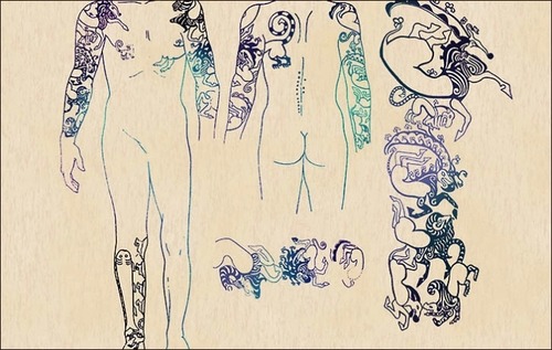
Style and design of tattoos may give a pretty good background idea (age, or cultural background of the decedent). Sometimes, if it is a customized piece of art, it may allow the investigators to find the artist as well. The archaeological discovery of the Siberian princess, Princess Ukok in 1993 has well demonstrated the great preservation of tattoo.

Altai Aborigines says the mummy of princess Ukok is their progenitor. The tattoo on her arm in fact has hidden some important message for mankind. They believe that she passed away voluntarily to protect the Earth from evil spirits. The tattoo covers from her shoulder to her hand, and only left arm tattoo was preserved. Left should was the canvas for a mythical animal, an antlered deer with the beak of a vulture. “The antlers are adorned with vulture heads; a similar head can be seen on the back of the animal, the body of which is twisted, followed by a sheep with its head thrown back. The mouth of a spotted panther with a long tail is seen at the legs of the sheep.” The clash between vultures and hoofed animals symbolize the conflict between the two worlds: a predator from the lower world against the middle world (symbolizes by a herbivorous animal).

(Source: Siberian Times)

(Source: Siberian Times)

Though researchers found later that the Siberian mummy was in fact died from breast cancer and self-meditated with cannabis, which crumbled the voluntary death speech from the aborigines, it supports the studies Byard quoted, saying tattoos in ancient times signifies the special social status of that tattooed person. And for Princess Ukok, she might not be a real princess, but was believed as a shaman because of the visions she saw after using cannabis.

** Recently, the face of Princess Ukok has been reconstructed. Thanks to the well developed facial reconstruction techniques. Click here to have a look of how she looked like.**

Resources:

Byard, W. Roger. 2013. “Tattoos: forensic considerations.” Forensic Sci Med Pathol (2013) 9:534-542.

Pravda.ru. 2011. “Tattoos of Princess of Altai conceal mankind’s biggest mysteries”

Siberian Times. 2012. “Siberian Princess Reveals Her 2500 Years Old Tattoos.”

United States Holocaust Memorial Museum. n.d. “ Tattoos and Numbers: The System of Identifying Prisoners at Auschwitz.” Holocaust Encyclopedia.

Crisis in Forensic Sciences, or Not?

Forensics is trusted by the authorities on convicting criminals for more than a century. Fingerprints, blood splatters, DNA, bit marks, shoe impressions, etc. are solid physical evidences that can associate a person with the crime, crime scene, or even put him on death row. Yet, forensic sciences are under strict scrutiny in the recent years. A lot of misjudgments are convicted because of wrong forensics.

The father back to the crime scene with Detective Stabler, the DA and the independent investigator/ professor for reenactment. (Source: Screen shot from Torch, Law & Order Season 11)

 If you are a crime drama fan like me, you may have watched every episode of Law & Order, CSI, or NCIS, to name a few. Yet, when talking about forensic scrutiny, Torch, an episode from Law & Order Season 11 particularly rings a bell.

 Let me walk you through the storyline:

The episode begins with an apartment caught a fire in the middle of the night, and two young girls died in a burning house. They were left in the house by their father, not because he did not try to save them. He did, yet the fire was too vigorous. He failed and burnt his hand. The (SENIOR, as he claimed himself) fire marshal concluded the fire was set by the father with incorrect assumptions he made. These assumptions were disarmed by an independent investigator/ professor, together with a real reenactment of the scenario—burning the DA’s house!

This episode alerts the public the danger of forensic sciences, or the danger of junk sciences. In fact, it does not only happen in drama but happens frequently in the reality.

In the documentary Forensics in Trial (watch here), it criticizes and investigates the uniqueness of fingerprints, the science-ness of bite marks. Jessica Gabel, a law scholar, commented that “forensic bit mark evidence is more art than science;” a lot of interpretation is needed indeed. In this case, should we still trust forensic sciences (are we, the forensics lovers living in our little fantasy)?

Both DNA and autopsy are hard sciences. The former is chemistry, and the latter is biology/ human anatomy. Are these two secure enough to convict crimes? Autopsy, for instance, may not be as accurate as it is. In the recent Michael Brown case, the autopsy is problematic. Shawn Parcells, the man that assisted in Michael Brown’s autopsy has his credentials questioned. He presented himself, according to Mail Online, as a pathologist even though he was not.

DNA, a well-known crime conviction technique, plays a critical role in cases such as the famous OJ Simpson case. Even the DNA evidence ties OJ Simpson to the crime scene, it took the jury only four hours to decide he is not guilty. Why? Because the plaintiff could not prove the evidences are beyond reasonable doubt in order to convict OJ Simpson. Part of the doubts is the legitimacy of evidence. The evidences had lost the chain of custody; police were accused for contaminating the crime scene, and plated evidence to frame Simpson. These all come together to show that even though the person was guilty, once the evidence did not speak the exact same story, no crime will be convicted.

By the same token, in 2012 a forensic chemist named Annie Dookhan had falsified thousands of drug tests. She had also mixed up evidence samples, fabricating results so as to be the most productive chemist in the lab, and lied about credentials.

All these examples just to illustrated one thing: it is not the science is false but the interpretation, and the person who interpreted the evidences are fractured. Quoting attorney McShane from Dookhan case, saying “a forensic scientist in a courtroom is that of a neutral scientists, not as part of the prosecution team…it’s the duty of a toxicologist to focus on the chemistry and the pharmacology not to concentrate on the conviction.”

Like any crime drama, people working in forensics and law enforcement do have a sense of justice, and what does it mean to each of them. One should not forget that being a good scientist, or even just law enforcement agent, professionalism and open-minded thinking are both critical.

The coolness of forensics is the accuracy of conviction. Mistaking one step would make killers walk free, while sending innocents to jails, even on death row.

Resources:

The OJ Simpson Case

Spargo, Chris. 2014. “The mystery of Shawn Parcells: How forensics ‘expert’ without a medical degree assisted in Michael Brown’s autopsy amid claims he stole a body and lost a brain.” Mail Online.

Trager, Rebecca. 2014. “Hard Questions After Litany Of Forensic Failures At US Labs.” Chemistry World.

Forensic Anthropology Internship Series Ep.4: Two Cases (and bone overgrown on the skull)

I really love walking into the morgue (I know it does not sound right), learning something new, and interacting with all these wonderful minds.

One of the autopsies I observed this time was about a woman found dead in her cabin on a cruise ship. She got her knee surgery before she boarded, and was prescribed antibiotics. When the Medical Examiner opened her up, she immediately found pulmonary fluid in her chest cavity (which could indicate infection). Later, she opened up her skull, and learned that her skull has a feature, Hyperostosis frontalis interna (HFI), which usually sees that on middle-aged women, sometimes men. The individuals with this feature are having a thicker frontal and occipital bone when compares with the parietal and temple bones. She also found a piece of little bone that is ossifying when she was removing her brain. Afterwards, she decided to dissect her knee too. When she opened the knee, it was totally infected. And the surgery apparently has removed her knee cap and replaced with an artificial one.

The bone from the surgery however never healed. Superficially, other than her surgical wound, she got few circular 3mm wide wounds (probably got from the infections).

****************************************************

More on Hyperostosis frontalis interna (HFI) (may choose to skip this and read on about the next case 🙂 )

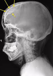
Hyperostosis frontalis interna (HFI) (Source: radiopedia.org)

Hyperostosis frontalis interna (HFI) is an independent yet pathological condition with an unknown etiology thus far. It refers to the manifestation of accretion of bone on the inner table of the frontal bone. Yet, this phenomenon of bone thickening on endocranial surface usually not extending beyond the coronal suture (Ruhli and Henneberg 2001). HFI was first described by Morgagni in 1769 and even with the great jump in medicine and pathology in almost three centuries, the origin and the detailed etiology are still unknown. Yet, with all the studies have done, HFI is most likely presence in post-menopausal women, which may link its presence to the hormone level disturbance in human body (Hajdu et al. 2009; Hershkovitz et al. 1999; Ruhli and Henneberg, 2001, May et al. 2010).

The origin of HFI still remains as a mystery till today. Hajdu et al. (2009) suggested that HFI could possibly link with unknown genetic predisposition, environment, hormones allocation and metabolic diseases. Specifically, HFI is clinically associated with Morgagni’s syndrome, or MSM—Morgagni-Stewart-Morel syndrome, which is a hormonal disorder that demonstrates symptoms like obesity, hirsutism and metal retardation (Barber et al., 1997; May et al., 2010).

Hershkovitz et al. (1999), May et al. (2010), and Ruhli and Henneberg (2001) purport that hormonal influences are key to understand the etiology of this phenomenon, especially about the hormonal regulation in females. Hershkovitz et al. (1999) quoted Ritcher (1939) that HFI under his study will disappear after passing of the endocrine imbalance of menopause. Ruhli and Henneberg (2001) hypothesized that because of the decrease of selective pressure has increased the metabolic rate due to the higher level of leptin (a 167 amino acid peptide coded by ob gene), which in turns may cause an increase of localized bone overgrowth (Rhuli and Henneberg, 2001). Given that leptin is strongly correlated to body mass index. Obesity, according to Haffner and Bauer (1993), is widely recognized as a major effect on bone density. This study suggests that however the adjustment for glucose and insulin concentrations does not explain the exact association between bone density and obesity, rather the bone density increased may be due to stress on the skeleton early in life, which lead to increase of bone formation, or to increase estrogen or androgen concentrations (Haffner and Bauer, 1993). Ruhli et al. (2004) particularly quoted Rudali (1968) changes of metabolism of female hormones in fat cells could link up HFI and adiposity. Other than leptin, Ruhli et al. (2004) suggested a possible microevolution of human sex steroids as the etiology of HFI.

The overgrowth bony tissue of HFI grows specifically on the inner plate of the frontal bone of the skull. It usually comes in the form of bilateral nodular, and only restricted to the area between the superior sagittal sinus medially and middle meningeal artery posteriorly (May et al., 2010). Yet, it is still unknown for frontal bone as the spot for this hormone target. Hershkovitz et al. (1999) suggested that one of clues might be the fact that HFI process “almost always begins in the middle one third of the frontal squama,” given that Morel (1930) considered that spot would be the original centers of ossification of the bone. Hormones such as estrogen may have trigger the primary center of ossification of the frontal bone to grow extra bones (Hershkovitz et al., 1999). HFI can be traced via dried skulls with naked eye inspection, radiographic studies and computerized tomography (CT) scan images. Yet, plain radiographies could be ineffective for detecting the early stages of HFI as opposed to naked eyes inspection as lesions in gauge can be distinguished) (Hajdu et al., 2010).

Various neurophysiologic, metabolic, radiologic studies performed and failed to suggest a direct linkage between HFI and behavioral disorders, rather suggesting the bone nodular lesions shown on x-ray are coincidence (Smith and Hemphill, 1956) Most of the cases of HFI are asymptomatic.

HFI are ethnicity-related and chronologically affected. Scholars like Hajdu and many others agreed that it seems that it is impossible to compare different populations and different centuries in order to get a general possible trend for now. Also, Raikos et al. (2011) pointed out that it is a rare case to find female with HFI in antiquity but males. Ruhli et al. (2004) compared the data collected before industrial revolution and those after and found resemble results as Raikos et al. (2011) but a big change for the data after industrial revolution. Ruhli et al. (2004) suggested the change of female life cycle through social developments and the longevity of modern population initiate the change. It is important to remember however, lower life span in historical times do not mean a lower prevalence of HFI (Ruhli and Henneberg, 2001).

***********************************************************

Back to the case…

The other male deceased was a 45-year man that survived from a four car accident April after he had a head surgery. The surgery, according to the Medical Examiner has taken out almost his right hemisphere of the skull to release pressure (which is called Trephination). He suspected that the accident caused a lot of internal bleeding, and the blood rushed all into the skull. The blood not only had increased the pressure inside the skull but also changed the shape of the brain—it presses the brain towards the left hemisphere of the brain.

The deceased has been in coma since, and stayed in the hospital. The coma did age him pretty fast (he looked like an old man by the time he was on the autopsy table) and took away lots of his nutrients.

ResearchBlogging.org

Hajdu, T., Fóthi, E., Bernert, Z., Molnár, E., Lovász, G., Ko˝vári, I., Köhler, K., & Marcsik, A. (2009). Appearance of hyperostosis frontalis interna in some osteoarcheological series from Hungary HOMO – Journal of Comparative Human Biology, 60 (3), 185-205 DOI: 10.1016/j.jchb.2008.07.004

Hershkovitz I, Greenwald C, Rothschild BM, Latimer B, Dutour O, Jellema LM, & Wish-Baratz S (1999). Hyperostosis frontalis interna: an anthropological perspective. American journal of physical anthropology, 109 (3), 303-25 PMID: 10407462

Raikos, A., Paraskevas, G., Yusuf, F., Kordali, P., Meditskou, S., Al-Haj, A., & Brand-Saberi, B. (2011). Etiopathogenesis of hyperostosis frontalis interna: A mystery still Annals of Anatomy – Anatomischer Anzeiger, 193 (5), 453-458 DOI: 10.1016/j.aanat.2011.05.004

Rühli, F., & Henneberg, M. (2002). Are hyperostosis frontalis interna and leptin linked? A hypothetical approach about hormonal influence on human microevolution Medical Hypotheses, 58 (5), 378-381 DOI: 10.1054/mehy.2001.1481

Forensic Anthropology Internship Series Ep.3: Bone Donation

Every beginning of a week in the Medical Examiner Office is the busiest, as the bodies came in throughout the weekend are all lined up for check-out check and autopsies on Monday. Also, a lot of parties going on, especially in Miami every weekend, young adults accidentally died from overdose, drunk drive and so on could happen more easily. Of one of the particular Monday, among the 5 cases that came in, there were two really remarkable ones.

Both of the cases are having young deceased individuals, with one female and one male respectively. The female was expired from a car accident, where the male was suffered nasal congestion suddenly and expired.

What really fascinating (I know this might sound inappropriate, apologize for that. Fascinating from the medical and forensic fashion,) is the stitches on both deceases’ limbs. We were told that some of their limb bones are taken out for bone donation. I have never thought about bone donation before (sorry for my ignorance). According to one of the morgue technicians, those fresh and healthy bones for “transplant” have to be removed within a day from the deceased since the heart stopped beating. After taking out the bones for donation, they would stuff towels back into the limbs and sew it up. The limbs do not look any differences, nor feel any differences when you first touch it. But when the morgue technician was trying to clean them lifted their limbs up and twisted it. The difference is huge!

Who can donate bones?

According to the Musculoskeletal Transplant Foundation, majority of bone donors were health and relatively young deceased who died in accidents, or sudden illness (heart attack or stroke). Screenings including medical histories and social histories (location of any high-risk behaviors for transmissible diseases will automatically eliminate from the pool for possible donation). Potential donors with histories of any condition that would affect long-term bone performance would also be excluded.

Bone donation counted as tissue donation. Unlike organ donation, tissue recipients do not have to match with the donors, as rejection is not the mainly concern. Organ donation donors are basically brain dead, and the donation process occurs when mechanical support that continue the organs to “live” for an extended short period of time after the death of the patient. Organ donation needs patience, as it requires matching blood type, medical status and so forth, whereas tissue donation only requires patients to be brain dead or cardiac death.

Also, for the lady who expired from the accident, she did not have any trauma in her body except for few broken ribs (which could possibly hurt her lungs), suspecting she might have been drink and drive. Her major blow was the blunt force trauma on her left posterior temple bone. The blow was so strong that the energy transmitted all across the occipital bone and reached the right temple bone, as well as fractured the base of the skull too. It gives me goose bumps. I hope that the blow did not kill her slow! The brain did not look abnormal except being bloody. The Medical Examiner says the blow could have done some traumas on the brain (yet we did not get to see the cutting of the brain today), but that amount of blood was sufficient to make her choke and swallow her own blood and caused aspiration on her lungs. Doctor says the blow should have her killed pretty quickly but may slow down due to the aspiration.

One personal thought:

I think if the government used the traumas and the fractures as warning for “Do not drink and drive” commercials would be working more effective than ordinary movie clips. This lady’s traumas definitely warn me more effectively to stay out of alcohol if I am driving.